| Return to the top page | |||
| Return | |||
| Projection patterns of single input/output axons of the cerebellum | |||
| We have been focused on this subject for a long time. How an axon of a single neurons extends, branches and terminates is one of the most fundamental information in understanding function of the neuron and the system which the neuron is involced in. There are climbing fiber and mossy fiber axons as inputs and Purkinje cell axons and axons of cerebellar nucleus neurons as outputs in the cerebellum. Projection patterns of these single axons are deeply related to the function of the cerebellum. | |||
| Reconstruction of a single mossy fiber axon (dorsal column nucleus neuson) | |||
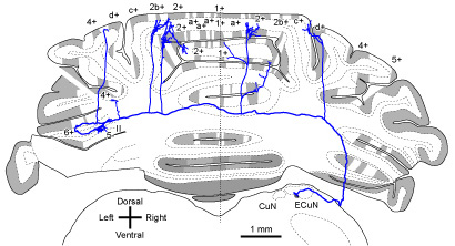 |
|||
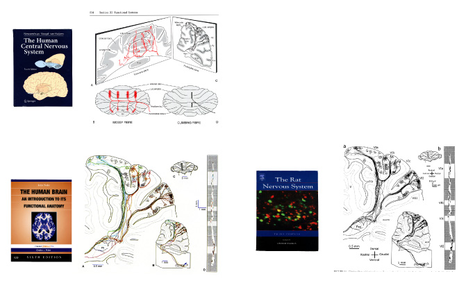
| Projection patterns of single climbing fiber axons and single mossy fiber
axons (Cited in textbooks) |
|||
 |
|||
| Director: Izumi Sugihara [Details] | |||
| Recent publications on this research subject Biswas MS, Luo Y, Sarpong GA, Sugihara I. (2019) Divergent projections of single pontocerebellar axons to multiple cerebellar lobules in the mouse. J Comp Neurol. doi: 10.1002/cne.24662. DOWNLOAD Luo Y, Patel RP, Sarpong GA, Sasamura K, Sugihara I. (2018) Single axonal morphology and termination to cerebellar aldolase C stripes characterize distinct spinocerebellar projection systems originating from the thoracic spinal cord in the mouse. J Comp Neurol 526(4):681-706. DOI: 10.1002/cne.24360. DOWNLOAD Luo Y, Sugihara I (2014) Cerebellar afferents originating from the medullary reticular formation that are different from mossy, climbing or monoaminergic fibers in the rat. Brain Research. DOWNLOAD. Sugihara I, Brown KM, Ascoli GA. (2013) New insights on vertebrate olivo-cerebellar climbing fibers from computerized morphological reconstructions. BioArchitecture 3(2):38-41. DOWNLOAD Fujita H, Sugihara, I (2013) Branching patterns of olivocerebellar axons in relation to the compartmental organization of the cerebellum. Front. Neural Circuits 7:3.DOWNLOAD Aoki H, Sugihara I (2012) Morphology of single olivocerebellar axons in the denervation-reinnervation model produced by subtotal lesion of the rat inferior olive. Brain Res. 1449(1) 24-37. DOWNLOAD Brown K, Sugihara I, Shinoda Y, Ascoli G. (2012) Digital morphometry of rat cerebellar climbing fibers reveals distinct branch and bouton types. J. Neurosci. 32(42) 14670-14686. DOWNLOAD. Cited in the cover illustration of the volume. Quy, P.N., Fujita, H., Sakamoto, Y., Na, J., Sugihara, I. (2011) Projection patterns of single mossy fiber axons originating from the dorsal column nuclei mapped on the aldolase C compartments in the rat cerebellar cortex. J. Comp. Neurol. 519(5): 874-899. DOWNLOAD Sugihara, I., Fujita, H. (2010) A computer-aided light microscopy system for three-dimensional reconstruction of axonal projections. In: A. Mendez-Vilas and J. Diaz eds. Microscopy: Science, Technology, Applications and Education, Microscopy Book Series No. 4., Badajoz: Formatex. pp. 813-819. DOWNLOAD Sugihara, I., Fujita, H., Na, J., Quy, P. N., Li, B. Y., Ikeda, D. (2009) Projection of reconstructed single Purkinje cell axons in relation to the cortical and nuclear aldolase C compartments of the rat cerebellum. J. Comp. Neurol. 512(2): 282-304. DOWNLOAD |
|||
| Return to the top page | |||
| Return |